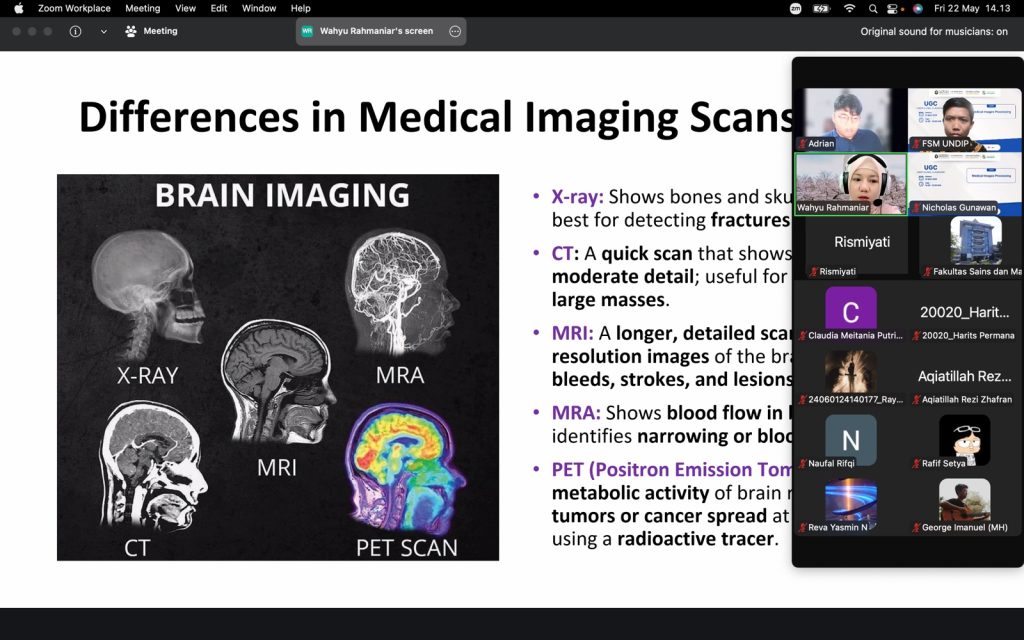
Semarang, 22 May 2026 — The Department of Informatics, Faculty of Science and Mathematics, Universitas Diponegoro, organized an UNDIP Global Classroom (UGC) session on Medical Images Processing on Friday, 22 May 2026. The online lecture was held via Zoom from 14.00 to 16.00 WIB, bringing together students, lecturers, and academic participants to discuss the growing role of image processing, artificial intelligence, and computer vision in modern healthcare.
The session featured Dr. Wahyu Rahmaniar, Assistant Professor at the Institute of Science Tokyo, Japan, as the invited speaker. Dr. Wahyu is an Indonesian researcher whose academic work focuses on artificial intelligence, medical imaging, and computer vision. Her profile shows extensive research experience in medical and biomedical image analysis, with publications indexed in international journals and conferences.
During the lecture, Dr. Wahyu introduced participants to the fundamental differences between several medical imaging modalities, including X-ray, CT scan, MRI, MRA, and PET scan. Each modality has different strengths: X-ray is commonly used to observe bones and detect fractures; CT scans provide fast cross-sectional imaging with moderate detail; MRI offers high-resolution visualization of soft tissues; MRA focuses on blood vessels and blood flow; while PET scans help identify metabolic activity, including cancer-related abnormalities.
Beyond introducing imaging techniques, the class also highlighted how medical image processing supports diagnosis, treatment planning, and clinical decision-making. Students were exposed to how computational methods can help detect abnormalities, segment organs or lesions, classify diseases, and improve the accuracy and efficiency of medical analysis.
A key part of the session was the discussion of Dr. Wahyu Rahmaniar’s research contributions. One example is her work on automated Cobb angle measurement for spinal curvature assessment. This research is important in the diagnosis and monitoring of scoliosis, where accurate measurement of spinal curvature is needed to support clinical evaluation. Her related publications include “Auto-CA: Automated Cobb Angle Measurement Based on Vertebrae Detection for Assessment of Spinal Curvature Deformity” in IEEE Transactions on Biomedical Engineering and “Automated Cobb Angle Measurement for Adolescent Idiopathic Scoliosis Using Convolutional Neural Network” in Diagnostics.
Another sample of her research is real-time segmentation and classification of calcaneal fractures in CT images. This work demonstrates how image processing and AI can assist in analyzing CT scans to detect and classify bone fractures more efficiently. Such research is highly relevant to medical image processing because it combines computer vision, classification algorithms, and clinical imaging data to support radiological interpretation.
Dr. Wahyu has also contributed to more recent research topics involving anomaly detection and collaborative medical imaging AI. Her listed publications include “Multi-AD: Cross-Domain Unsupervised Anomaly Detection for Medical and Industrial Applications” in Pattern Recognition and “Future of the Medical World: Collaborative Medical Imaging AI with Federated Learning” in IEEE Consumer Electronics Magazine. These studies show how AI can be applied not only to detect unusual patterns in images, but also to support privacy-aware collaboration between medical institutions through federated learning.
In addition, her conference research includes medical imaging topics such as lesion detection and classification in digital breast tomosynthesis, segmentation of rare soft-tissue sarcomas in multi-sequence MRI, and lung nodule classification using a compact MTANN model. These examples gave students a broader view of how medical image processing can be applied to cancer detection, MRI analysis, and lung disease classification.
The lecture was moderated by Satriawan Rasyid P., S.Kom., M.Cs., from the Faculty of Science and Mathematics, Universitas Diponegoro. Through the discussion, participants gained insight into both the theoretical foundations and real-world applications of medical image processing, especially in relation to artificial intelligence and computer vision.
This UGC activity reflects Universitas Diponegoro’s commitment to strengthening international academic exposure and providing students with global learning opportunities. By inviting an international researcher with strong expertise in AI-based medical imaging, the session encouraged students to understand how informatics can contribute directly to healthcare innovation.
The Medical Images Processing class is expected to inspire students to explore interdisciplinary research that connects computer science, artificial intelligence, and medicine. It also emphasized that future healthcare systems will increasingly rely on intelligent technologies capable of assisting doctors, improving diagnostic accuracy, and making medical services more efficient and accessible.